About 5% of the female population suffers from some type of uterine malformation due to genetic problems, intrauterine infections during pregnancy or radiation exposure during embryonic development.
Most of these malformations can affect the ability to conceive or give birth, although some of them can be corrected with surgery.
Before we explain the types of pathological uterus that can be treated by surgery, we will tell you more about the uterus and its role during pregnancy.
Function and layers of the uterus
As you probably already know, the function of the uterus, as an essential part of the female reproductive system, is to provide the embryo with the right conditions and environment for its development.
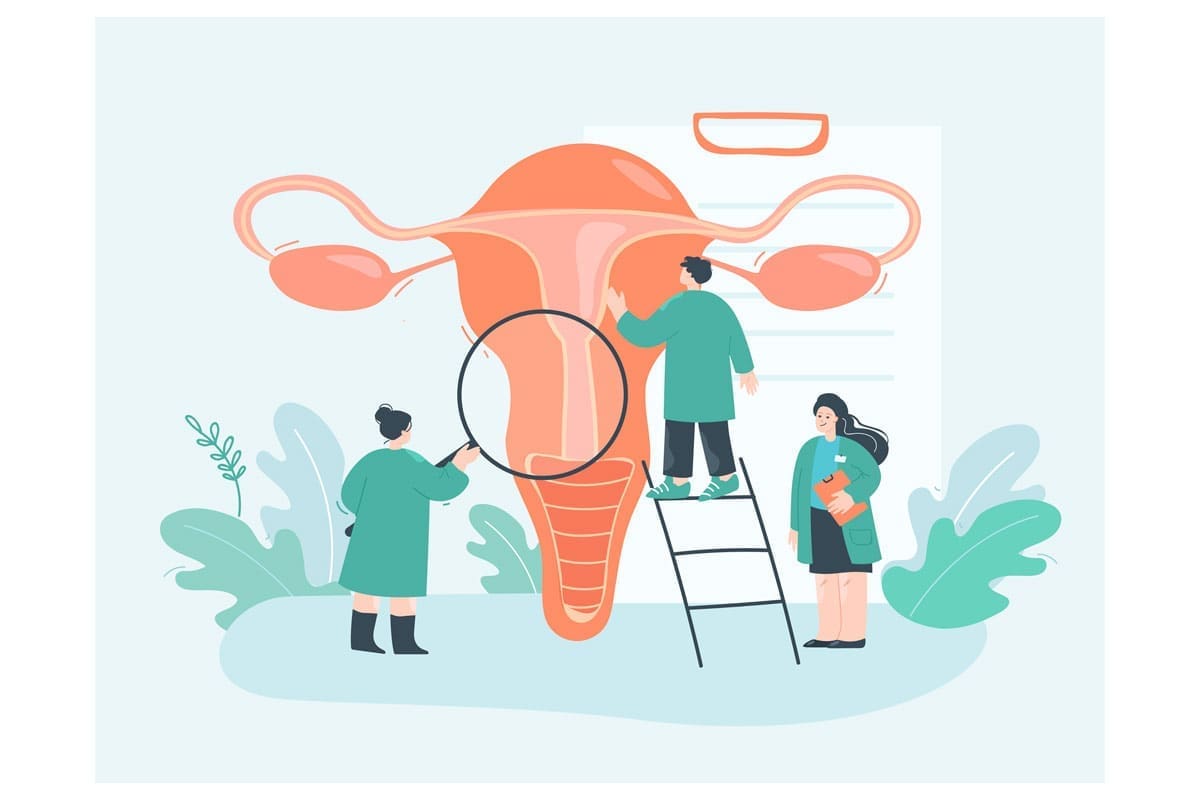
At a physiological level, the uterus is a hollow muscular organ, shaped like an inverted triangle and located in the pelvic cavity (in front of the rectum and behind the bladder).
The walls that make up the structure of the uterus are made up of three layers:
- The endometrium or inner layer.
Its receptivity (composition, thickness, uniformity…) will determine whether the embryo can nest and continue its development, once the embryo transfer has taken place.
This layer will vary throughout the menstrual cycle and, when pregnancy does not occur, it is shed and expelled in the form of menstruation.
- The myometrium or intermediate layer of the uterine wall (located between the perimetrium and the endometrium).
It is the thickest part and is formed by muscular tissue that will expand during pregnancy so that the uterine cavity can carry the baby.
During childbirth, the function of the myometrium will be key; it is through the contraction and expansion of this muscle that the mother will be able to give birth to the baby by expelling it through the vagina (in the case of a natural childbirth).
- The perimetrium or external layer.
Also called “serosa“. This is a thin layer of tissue that facilitates contact between the uterus and the other surrounding organs.
Parts of the uterus
On the other hand, the three parts into which this reproductive organ is divided are:
- The body.
The body is the widest part and where the development of the embryo takes place.
Its upper part, pear-shaped, forms the uterine fundus, on whose sides the fallopian tubes open.
- The cervix.
The narrowest and lowest part of the uterus.
It can be defined as a firm and smooth but also rounded structure. It is inserted into the vagina and it is through this opening that menstrual secretions (endometrial cells) are expelled.
- The isthmus.
The small circular base that joins the two parts already mentioned: the body and the cervix.
Uterine malformations and reparative surgeries
According to the European Society of Human Reproduction and Embryology (ESHRE) there are 7 main groups of uterus. Each of them, in addition, includes different subtypes that you can learn about in the article: “Types of uterine malformations: classification and main characteristics”.
However, not all uteri can be treated surgically at the reproductive level.
The main function of uterine interventions in reproductive surgery will be to repair the organ in order to restore fertility. Thus, in most cases, the woman will be able to have children through assisted reproduction treatment or even, on certain occasions, naturally.
However, when we speak of uterine malformations, in spite of the numerous advances that are made every year in reproductive medicine, there are uteruses with a morphology so different from what we consider “normal” in gynecology, that pregnancy will not even be possible to happen through surgery.
The uteri that we still cannot treat by means of reproductive surgery are:
- Infantile uterus
- Bicorneal or bicorporeal uterus.
- Unicornuate or hemiuterus uterus
- Dysplastic or aplastic uterus
On a positive note, these types of uterus represent only 15% of uterine malformations. The remaining percentage can be treated surgically by means of minimally invasive interventions.
This 85% of malformations would be composed of:
- T-shaped uterus
- Partial septum uterus
- Complete septate uterus
Types of reproductive surgeries
First of all, the T-shaped uterus is characterized because, although externally it has a similar shape and size to a normal uterus, internally it is much smaller. Its name is due precisely to the morphology of its internal cavity.
One of the main anomalies of this type of uterus is that its walls are much thicker than usual.
In this type of case, our team of surgeons will opt for the elimination of the tissue excess in the lateral walls of the uterus through hysteroscopy. This way, we will enlarge the internal cavity, giving it an inverted triangle shape similar to a normal uterus.
On the other hand, the partial septum and complete septum uteri are characterized because, although both again have a conventional external structure, the inner part has a central wall or septum. This septum grows from the bottom of the uterus (upper part) dividing the uterine cavity into two parts.
The intervention that we will normally perform to repair this type of malformation will have a similar approach to that of the T-shaped uterus. That is to say, the objective will also be to remove the excess tissue by hysteroscopy.
However, the difference between the two surgeries lies in the fact that, in the latter case, the tissue to be removed will be that of the central wall. This way, we will be able to create a uterine cavity with a normal shape and size.
We are fertility specialists
Uterine malformations and the different types of uterus associated with them can negatively influence female fertility, preventing pregnancy.
The main problem is that, in most cases, women are completely unaware that they suffer from this pathology. This is because many of the malformations we have mentioned may not have any obvious side effects until the moment of seeking pregnancy. It is then when infertility manifests itself and we need the help of a gynecologist who is an expert in the diagnosis of uterine pathologies.
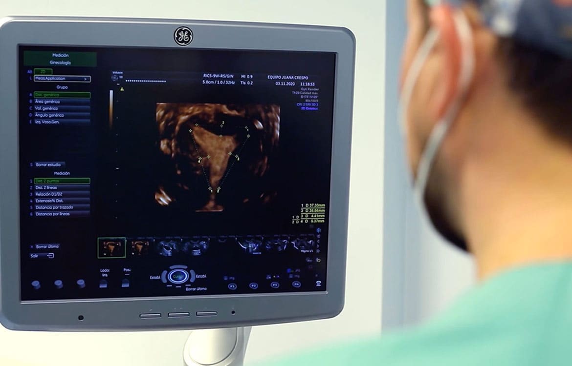
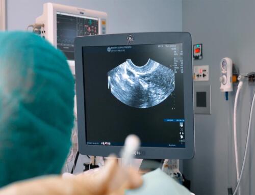
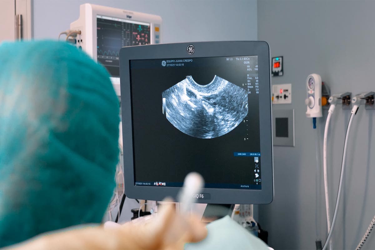
In Equipo Juana Crespo we are specialists in the detection of infertility causes because, as we usually say, infertility always has a reason. Some tests such as ultrasound, hysterosalpingography and diagnostic hysteroscopy allow us to detect malformations in the uterus depending on the particular abnormality and reproductive history of each woman.
If you have questions about your particular case and would like more information, please do not hesitate to contact us!


