.
In a biological sense, an embryo is an organism that is in the initial stages of its development.
In the case of humans, the embryo is called so, from the time the male (sperm) and female (egg) gametes merge until the eighth week of gestation.
Embryo quality in fertility treatments
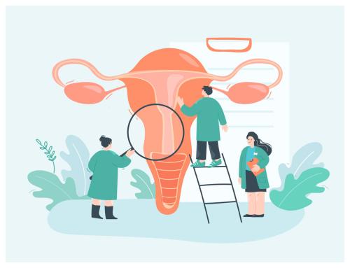
In previous articles we have talked about the importance of certain pathologies in fertility, such as endometriosis, adenomyosis, hydrosalpinx ….. Disorders and diseases that, in most cases, we will have to treat previously to increase the chances of success in assisted reproduction treatments.
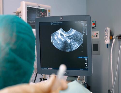
To these pathologies are added factors such as age, the state and morphology of the uterus, the adequate endometrial receptivity, etc.
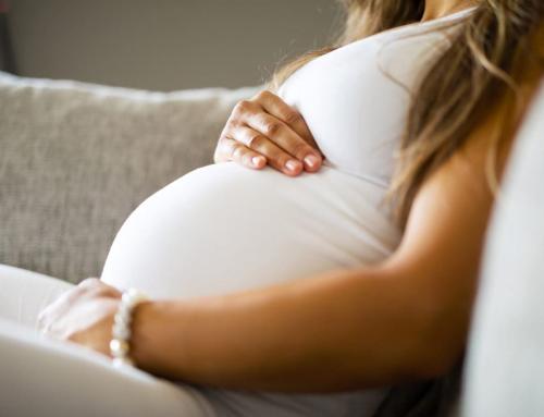
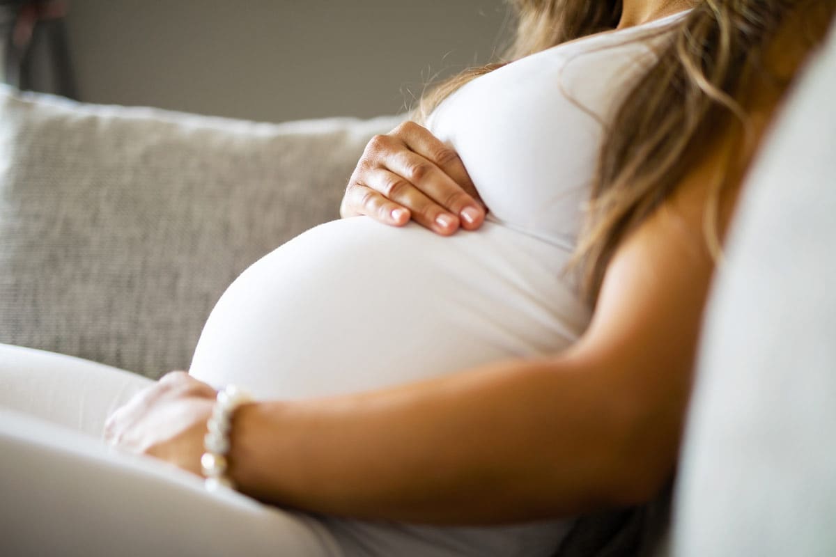
Therefore, although one of the main objectives of assisted reproduction is to ensure the formation of healthy embryos in order to achieve pregnancy, embryo quality is by no means the only aspect to be taken into account.
How the embryo is developed in an assisted reproduction treatment
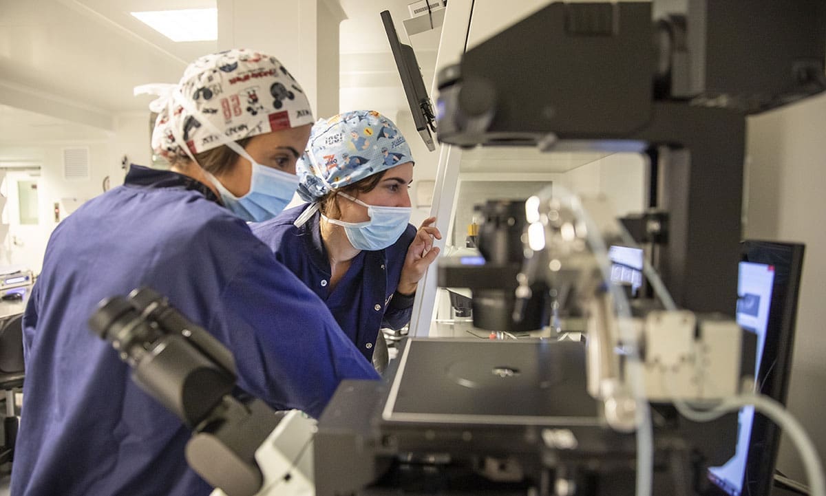
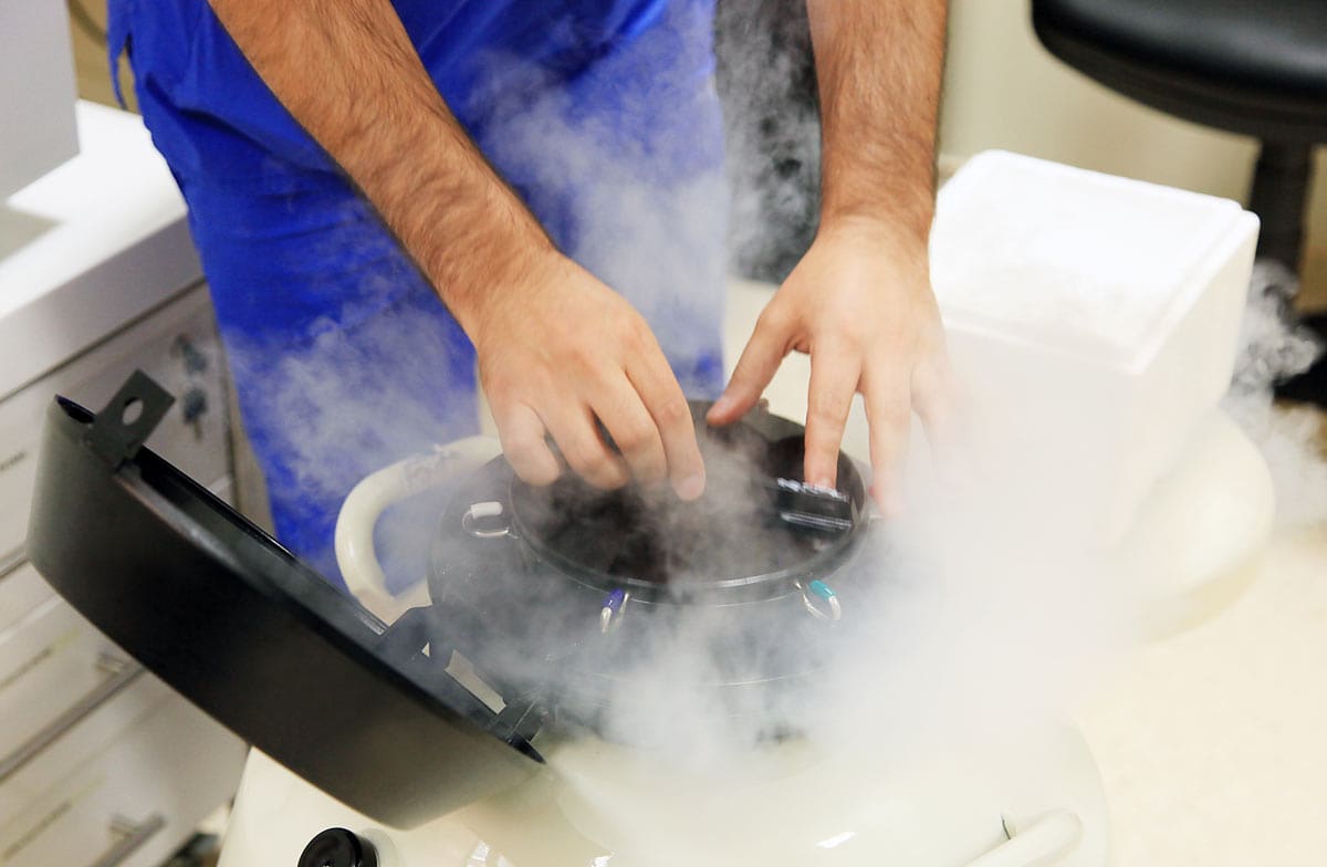
The creation of the embryo in IVF treatments is performed by extracting the male and female gametes. Once obtained and treated individually in the laboratory, fertilization is performed.
For this, we will use techniques such as ICSI or intracytoplasmic injection, which consists of perforating the cortex of the egg by means of a microinjection. Through it, we will deposit the sperm into the cytoplasm, thus facilitating fertilization to take place.
ICSI can be performed with the couple’s own sperm and egg, with donor sperm and the couple’s own egg, with the couple’s own sperm and egg donation or, lastly, with donor egg and sperm. This last option is what is known as embryo adoption.
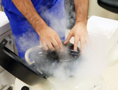
Normally, once the embryo has reached the stage of development known as blastocyst (5-6 days after fertilization), the embryo transfer will take place.
What are the stages of an embryonic development?
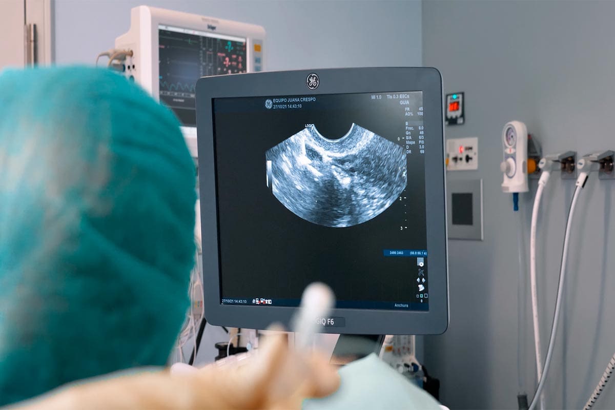
The process of embryonic development is called embryogenesis. In assisted reproduction treatments, the first signs of embryonic development take place in the laboratory, during embryo culture, when the egg is fertilized and becomes a zygote.
- Pre-embryonic period: what an embryo looks like during its first weeks of life
The pre-embryonic period, known as the germinal phase, is the first and shortest phase of embryonic development. It lasts from fertilization to the second week of the embryo’s life.
In this phase of embryo development, the zygote repeatedly self-replicates. This division gives rise first to the morula and then to the blastula.
When the embryo is in the morula stage (around the fourth day after gestation), it is a mass of undifferentiated cells named for its rounded, blackberry-shaped appearance.
Around the fifth day of its development, the embryo begins to grow a different type of cells, the blastomeres, which multiply and begin to organize themselves into three differentiated layers.
At this stage of development the embryo acquires the name of blastocyst. During this stage the embryo has a central cavity (blastocele) and two distinct cellular regions: the trophoblast or trophectoderm (precursor of the placenta and extra-embryonic tissues) and the inner cell mass (ICM, precursor of the embryonic tissue).
- Embryonic period: what the embryo looks like until it becomes a fetus
From the third week of gestation, gastrulation occurs, a process by which the embryo acquires its three germ layers.
At this time, the embryo has a rounded shape and the blastula cell layers have completely differentiated, giving rise to three structures: the ectoderm, the mesoderm and the endoderm.
These structures are the origin of the baby and all its tissues and organs, thanks to a process known as organogenesis.
- The ectoderm is the outermost layer and is composed, in turn, of three structures: the external ectoderm, the neural crest and the neural tube. The ectoderm is the origin of the nervous system and the epidermis.
- The mesoderm will originate in the bones, muscles, kidneys, circulatory system (blood and lymphatic cells), cartilage, connective tissue, adipose tissue and reproductive system.
- The endoderm, the last layer to develop and also the innermost, will be the origin of the lungs and airways, digestive system, liver, pancreas and urinary bladder. The endoderm also forms the endothelium of the endocrine glands or the thyroid.
The embryonic phase lasts eight weeks since gestation. The embryo’s appearance changes a lot during this time, as it grows about one millimeter each day. In addition, its rounded shape elongates until it forms a silhouette similar to that of a baby.
Thanks to technology, we can now see what an embryo looks like and differentiate its parts. We also know that in the embryonic phase the future baby acquires many of its basic physical features, both internally and externally: the head, face, limbs, body systems and internal organs begin to develop, and the first movements appear.
During the eighth week of gestation, at the end of the embryonic period, the embryo measures between 4 and 5 centimeters and weighs about 9 grams.
What is the difference between embryo and fetus?
Embryo and fetus are concepts that tend to be confused. Therefore, from Equipo Juana Crespo we will try to explain their differences in a simple way.
The biological stages from fertilization to the development of the fetus are known as “Intrauterine development phases” and they are three: pre-embryonic period, embryonic period and fetal period. The term embryo will be used only for the first two, which we have discussed in this post.
The embryo receives this name from implantation until the eighth week of gestation; after that, it will be called fetus.
The differences between the embryo and the fetus are related to the size and the degree of cellular, morphological and physiological development. To begin with, during the embryonic phase, the body proportion of the embryo is different, since the head has a much larger volume than the rest of the body, whereas in the fetal phase, the rest of the fetal body will increase in size and its shapes will balance out to form its definitive silhouette.
In addition, the fetus has a much higher level of cellular specialization than the embryo, since from the ninth week of gestation the organs are being created and will begin to function.
Are you interested in having a personalized diagnosis of your fertility or are you looking for information regarding our assisted reproduction treatments? Write to us, call us or visit us.
In a biological sense, an embryo is an organism that is in the initial stages of its development.
In the case of humans, the embryo is called so, from the time the male (sperm) and female (egg) gametes merge until the eighth week of gestation.
Embryo quality in fertility treatments
In previous articles we have talked about the importance of certain pathologies in fertility, such as endometriosis, adenomyosis, hydrosalpinx ….. Disorders and diseases that, in most cases, we will have to treat previously to increase the chances of success in assisted reproduction treatments.
To these pathologies are added factors such as age, the state and morphology of the uterus, the adequate endometrial receptivity, etc.
Therefore, although one of the main objectives of assisted reproduction is to ensure the formation of healthy embryos in order to achieve pregnancy, embryo quality is by no means the only aspect to be taken into account.
How the embryo is developed in an assisted reproduction treatment
The creation of the embryo in IVF treatments is performed by extracting the male and female gametes. Once obtained and treated individually in the laboratory, fertilization is performed.
For this, we will use techniques such as ICSI or intracytoplasmic injection, which consists of perforating the cortex of the egg by means of a microinjection. Through it, we will deposit the sperm into the cytoplasm, thus facilitating fertilization to take place.
ICSI can be performed with the couple’s own sperm and egg, with donor sperm and the couple’s own egg, with the couple’s own sperm and egg donation or, lastly, with donor egg and sperm. This last option is what is known as embryo adoption.
Normally, once the embryo has reached the stage of development known as blastocyst (5-6 days after fertilization), the embryo transfer will take place.
What are the stages of an embryonic development?
The process of embryonic development is called embryogenesis. In assisted reproduction treatments, the first signs of embryonic development take place in the laboratory, during embryo culture, when the egg is fertilized and becomes a zygote.
- Pre-embryonic period: what an embryo looks like during its first weeks of life
The pre-embryonic period, known as the germinal phase, is the first and shortest phase of embryonic development. It lasts from fertilization to the second week of the embryo’s life.
In this phase of embryo development, the zygote repeatedly self-replicates. This division gives rise first to the morula and then to the blastula.
When the embryo is in the morula stage (around the fourth day after gestation), it is a mass of undifferentiated cells named for its rounded, blackberry-shaped appearance.
Around the fifth day of its development, the embryo begins to grow a different type of cells, the blastomeres, which multiply and begin to organize themselves into three differentiated layers.
At this stage of development the embryo acquires the name of blastocyst. During this stage the embryo has a central cavity (blastocele) and two distinct cellular regions: the trophoblast or trophectoderm (precursor of the placenta and extra-embryonic tissues) and the inner cell mass (ICM, precursor of the embryonic tissue).
- Embryonic period: what the embryo looks like until it becomes a fetus
From the third week of gestation, gastrulation occurs, a process by which the embryo acquires its three germ layers.
At this time, the embryo has a rounded shape and the blastula cell layers have completely differentiated, giving rise to three structures: the ectoderm, the mesoderm and the endoderm.
These structures are the origin of the baby and all its tissues and organs, thanks to a process known as organogenesis.
- The ectoderm is the outermost layer and is composed, in turn, of three structures: the external ectoderm, the neural crest and the neural tube. The ectoderm is the origin of the nervous system and the epidermis.
- The mesoderm will originate in the bones, muscles, kidneys, circulatory system (blood and lymphatic cells), cartilage, connective tissue, adipose tissue and reproductive system.
- The endoderm, the last layer to develop and also the innermost, will be the origin of the lungs and airways, digestive system, liver, pancreas and urinary bladder. The endoderm also forms the endothelium of the endocrine glands or the thyroid.
The embryonic phase lasts eight weeks since gestation. The embryo’s appearance changes a lot during this time, as it grows about one millimeter each day. In addition, its rounded shape elongates until it forms a silhouette similar to that of a baby.
Thanks to technology, we can now see what an embryo looks like and differentiate its parts. We also know that in the embryonic phase the future baby acquires many of its basic physical features, both internally and externally: the head, face, limbs, body systems and internal organs begin to develop, and the first movements appear.
During the eighth week of gestation, at the end of the embryonic period, the embryo measures between 4 and 5 centimeters and weighs about 9 grams.
What is the difference between embryo and fetus?
Embryo and fetus are concepts that tend to be confused. Therefore, from Equipo Juana Crespo we will try to explain their differences in a simple way.
The biological stages from fertilization to the development of the fetus are known as “Intrauterine development phases” and they are three: pre-embryonic period, embryonic period and fetal period. The term embryo will be used only for the first two, which we have discussed in this post.
The embryo receives this name from implantation until the eighth week of gestation; after that, it will be called fetus.
The differences between the embryo and the fetus are related to the size and the degree of cellular, morphological and physiological development. To begin with, during the embryonic phase, the body proportion of the embryo is different, since the head has a much larger volume than the rest of the body, whereas in the fetal phase, the rest of the fetal body will increase in size and its shapes will balance out to form its definitive silhouette.
In addition, the fetus has a much higher level of cellular specialization than the embryo, since from the ninth week of gestation the organs are being created and will begin to function.
Are you interested in having a personalized diagnosis of your fertility or are you looking for information regarding our assisted reproduction treatments? Write to us, call us or visit us.


